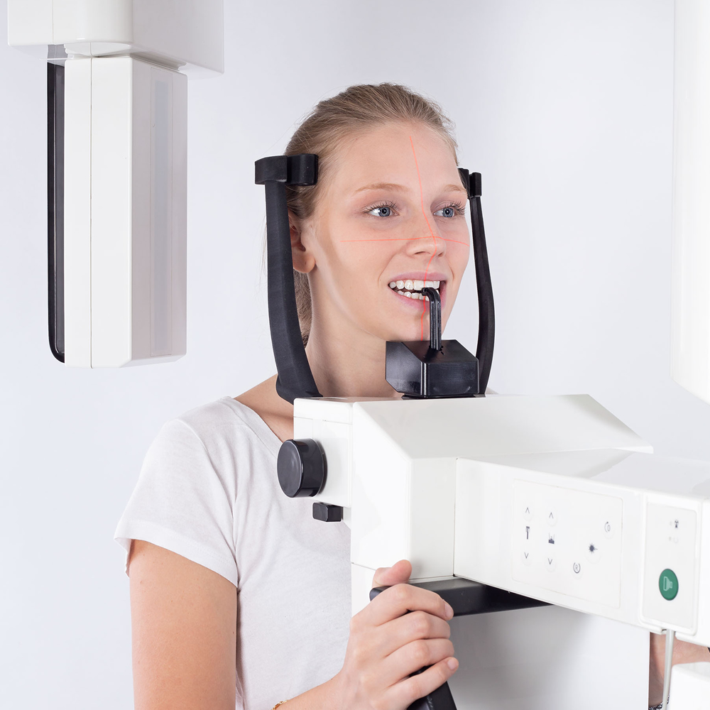
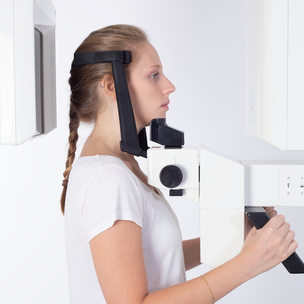
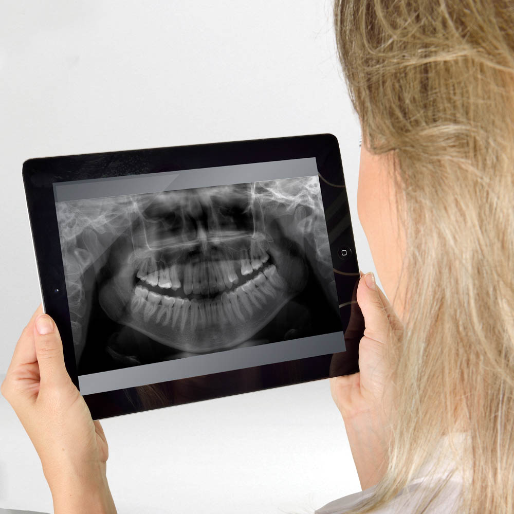
Eagle 3D PAN
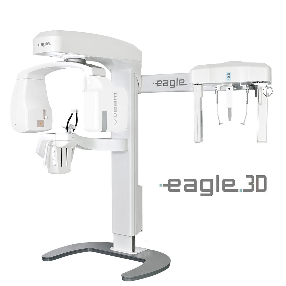
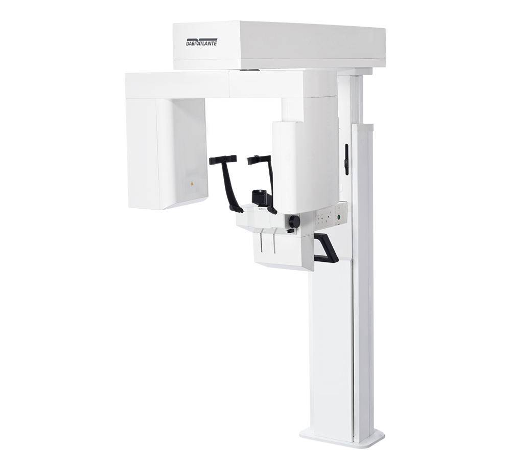
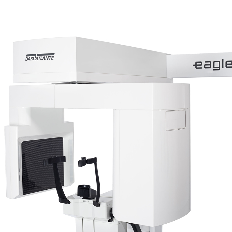
The Eagle 3D PAN is an equipment that gathers together all functions of the panoramic profile with the odontological tomograph. The 3D Module offers the exclusive VBeam technology with up to 8 field of view (FOV) volumes, from 5x5 to 23x16.
The exclusive VBeam technology - Variable Cone Beam - developed by Dabi Atlante, ensures high definition for images with up to 5x5, 6x8 and 8x8 FOV, as well as allowing capture of larger images (up to 23x16).
The Eagle 3D allows for upgrade to the more complete Pan/Ceph 3D version with FOV up to 23x16, making it a 3-in-1 equipment.
There are currently 3 versions of the Eagle 3D PAN for sale: with 3 FOV sizes (5x5-6x8-8-8), 4 sizes (5x5-6x8-8x8-8x12) or 8 sizes (5x5-6x8-8x8-8x12-8x16-13x16-18x16-23x16). Upgrade is also possible among the versions.
The Eagle line offers high image quality, easy handling, easiness in positioning the patient and gathers state-of-the-art world technology for all functions. The high frequency X-ray generator reduces unnecessary patient exposure. The micro-processed high speed system and accurate mechanics allow for perfect execution and repeatability in 2D and 3D images.
Eagle Technology
Eagle V-Beam
Variable Cone-Beam, an exclusive technology developed by Dabi Atlante, guarantees high definition in images with FOV of 5x5, 6x8 and 8x8, and also allows for capturing images of larger dimensions (up to 23x16).
8 FOV
5x5 - 6x8 –8x8 - 8x12 - 8x16 – 13x16 - 18x16 - 23x16.
80µm resolution
The Eagle 3D has different resolutions with Isotropic Voxel between 80 and 500 µm.
Automatic Processing
Regardless of the chosen profile, the images are automatically processed and displayed within the sharpen, brightness and contrast parameters previously determined, giving agility to the workflow and standardization of the exams.
Easily Patient Positioning
The adjustable height of between 1,20m and 2m, for the frontal positioning of the patient in relation to the operator, tooth clamp with swivel brake, chin support, temple support and three laser beams, guaratee precise positioning of the patient.
On Demand
One of the most renowned software for tomographic reports in the world. The software offers a friendly interface, availability of resources/functions, speed in processing, security, and total display/management of the generated images.
DICOM SERVER
The Eagle DICOM Send software tool permits the instant transmission of images generated by Eagle to storage and sharing systems in physically separate locations; an extremely useful resource for clinical networks that can decentralize image acquisition and centralize reports.
| Supply Voltage | 110/127/220 or 240V |
|---|---|
| Power Consumption | 1,25 KVA |
| Operating Frequency | 50 or 60 kHz |
| Generator Type | High Frequency |
| Focal point | 0,5mm |
| Voltage tube | 60-85kV |
| Tube current | 4-8mA |
| Image detector | CCD/CMOS |
| Pixel size | 108 µm (Pan e Ceph)/ 100 µm (3D) |
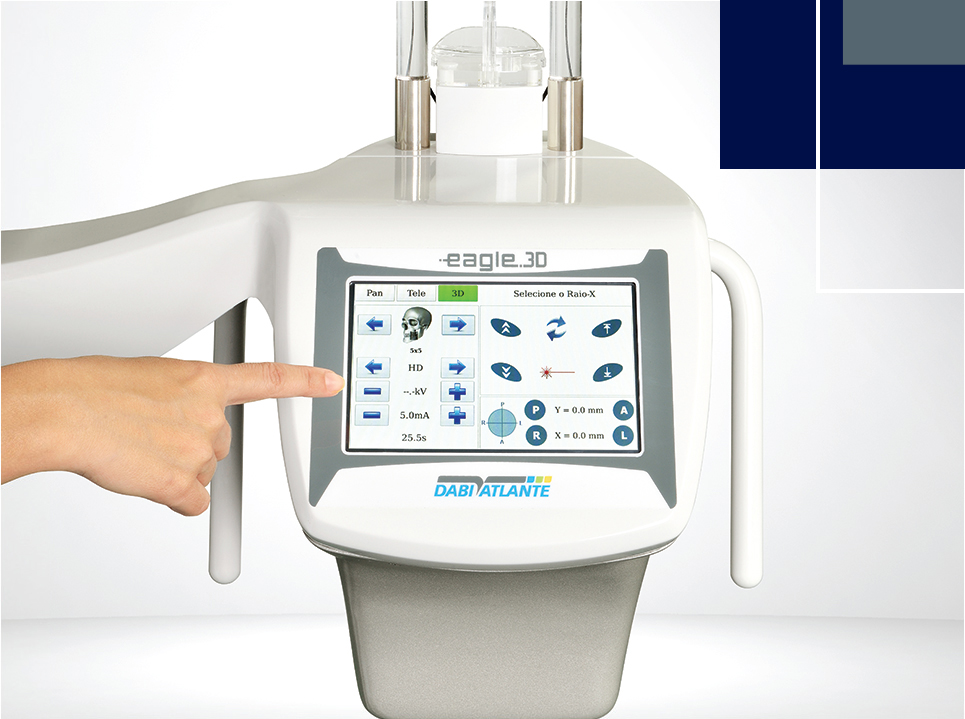
| Display | Touchscreen |
| Panoramic Profile | Yes |
| Cephalometric Profile | Optinoal |
| Tomography (3D) Profile | Yes |
| Sensor quantity | 1 sensor (2D) or 1 sensor (3D) |
| Base | Optional |
| Acessible to wheelchair user | Yes |
| PAN | |
| Exposure (time/current): | |
| PAN - Standard | 14s - 8mA |
| Improved Ortogonally | 14s - 8mA |
| PAN - low dosage | 13s - 6,3mA |
| PAN - children | 11,5s - 6,3 mA |
| Bitewing | 7,6s - 8mA |
| Improved Ortogonally Bitewing | 7,6s - 8mA |
| Maxillary Sinus | 8s - 8mA |
| ATM | 10s - 8mA |
| Average expasion | 1:1,16 |
| INFORMAÇÕES RADIOLÓGICAS - TOMOGRAFIA | |
| FOV | Configuração 1 - 5x5|6x8|8x8| Configuração 2 - 5x5|6x8|8x8|8x12| Configuração 3 - 5x5|6x8|8x8|8x12|8x16|13x16|18x16|23x16 |
| Voxel | 80 – 500 µm |
| PC SPECIFICATIONS | |
| Operating System | Windows 7 Professional - 64Bits Windows 8 Professional - 64Bits Windows 8.1 Professional - 64Bits Windows 10 Professional - 64Bits |
| CPU | Intel Core i7 4.0 GHz or higher |
| RAM | 16GB DDR2 SDRAM |
| HDD | 1TB or higher |
| PCI | PCI Express (PCIe) slot |
| NIC | Gigabit Ethernet dedicated |
| Video | NVIDIA GEFORCE GTX 1060 6GB or higher |
| USB Connector | 3x USB 2.0 |
| Monitor | 1920 x 1080 - Full HD |
| Power Suply | 400W RMS with supplementary power connectors PCI Express compatible with video board |
| MINIMUM DIMENSIONS OF THE ROOM | |
| Width | 1,8m |
| Length | 1,9m (Without cephalometric arm) 2,5m (with cephalometric arm) |
| Height | 2,5m |
2D Radiographic exams - Pan Profiles

Standard pan
This exposure has constant vertical magnification in the region of the dental arch, provides great width to the cutting plane and prioritises uniformity of the exposure throughout the image.
T.M.J
This double exposure shows the region of the dental condyles with the mouth open and closed in the same image.
Maillary sinus
This exposure focuses on the maxillary sinus region.
Improved ortogonally*
This exposure contains the standard pan profile with improved orthogonality for the beams in relation to angles thus providing for reduced reassembly of teeth on the exposure.
Low dosage*
This exposure corresponds to the standard panprofile and is perfomed ina fast exporure and using a lower anode current (mA). The image quality is still suitable for diagnosis although the dosage is less tha that for standard pan exposure with great image quality.
Children pan
This exposure features a reduced profile, 15% less tha for the standard pan.
Bitewing
This exposure has an image profile type for the bitewing area of the pre-molars and molars, including part of the maxilla, mandible and mandibular ramus.
Improved ortogonally bitewing*
This exposure contains the bitewing profile with improved orthogonality for the beams in relation to angles thus providing for less reassembly of teeth on the exposure.2D Radiographic exams - Ceph Profiles

Digital ceph profile
With this profile it is possible to obtain the following digital images:PA; Posterior-Anterior
AP; Anterior-Posterior
45 Degrees
Lateral
Carpal
PA de Water´s View
Axial Basal - Hirtz View.

Low dosage digital ceph profile*
With this profile it is possible to obtain a lateral display with a reduced exposure area and thus a reduced dosage for the patient.*Available only on the digital version.
8 volumes for tomographic acquisition.

5x5 ENDO
Small FOV optimized for local diagnostics, such as single implant planning, extraction of the 3rd molar, and endodontic prcedure. It keeps the patient's exposure dose to a significanttly reduced level.Mantém a dose de exposição do paciente em um nível sensivelmente reduzido.
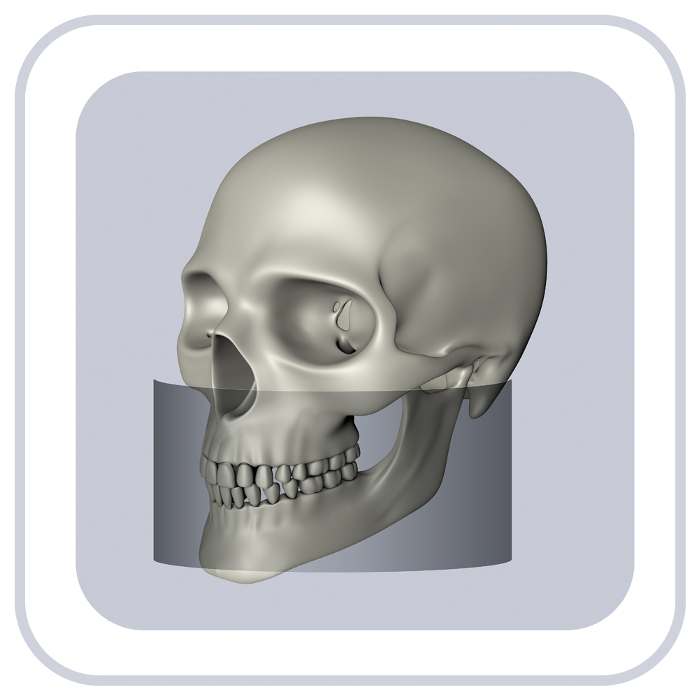
8x16 - EXTENDED JAW
Allow for display of the mandible, maxilla, nasal passages, sinus, and both TMJs in the same image.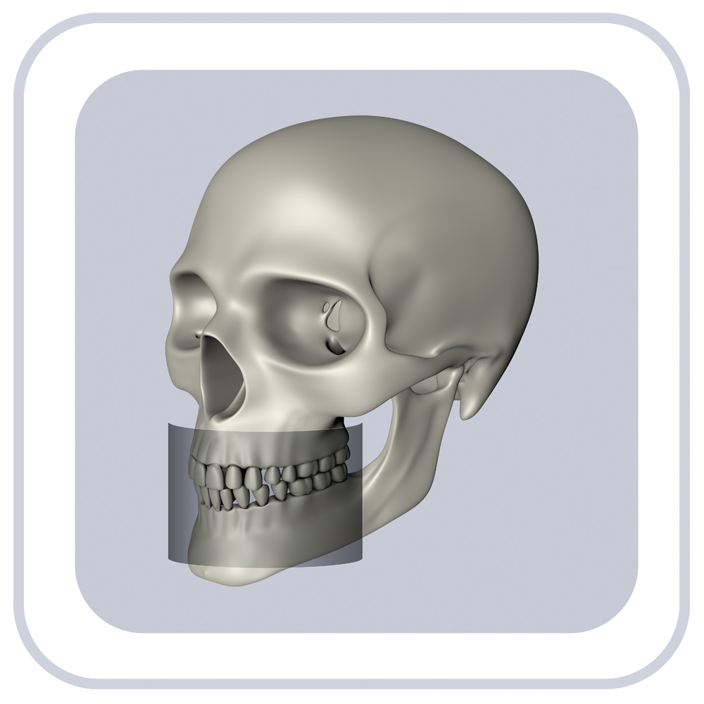
6x8 UPPER/LOWER JAW
Allow for display of the maxilla or mandible.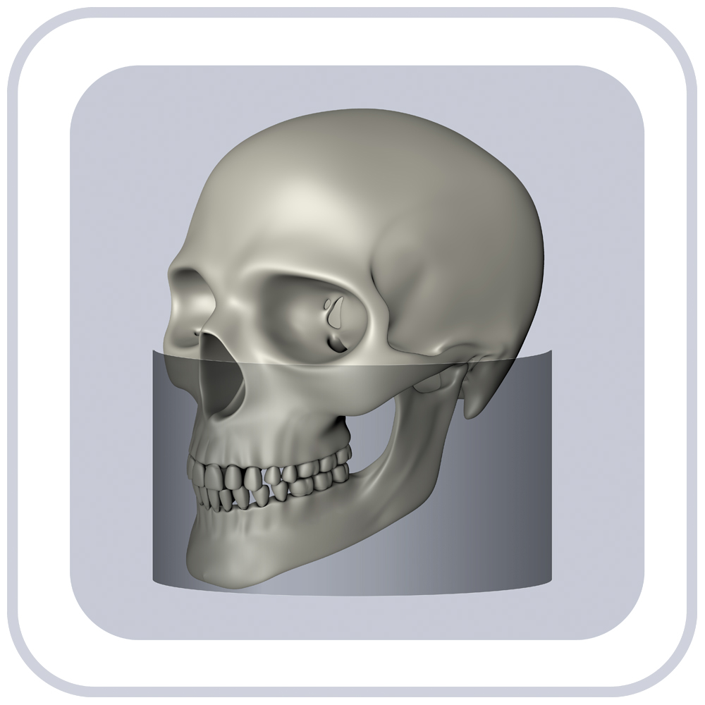
13x16* - SKULL
ideal for orthodontics, it allows for diagnostics on the whole maxillofacial region.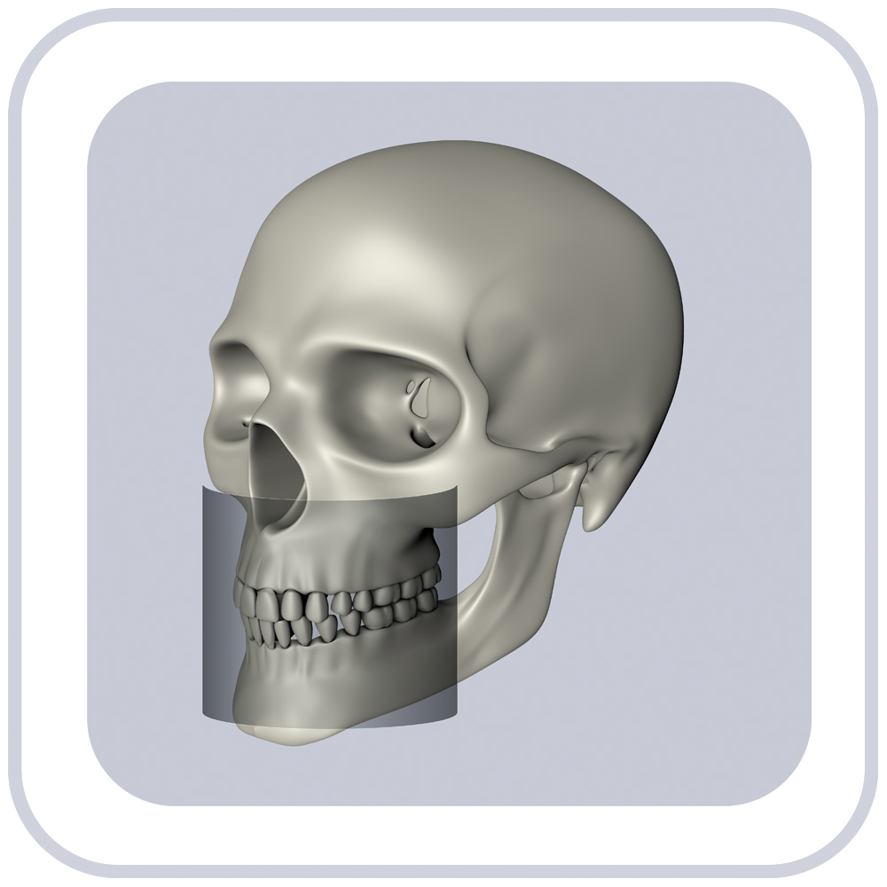
8x8 - FULL JAW
Covers the whole jaw, including mandible, maxilla, and part of the maxillary sinus.
18x16* - SKULL
Ideal for orthodontics, its allow for diagnostics on the whole maxillofacil region.
8x12 - FULL JAW
Covers the whole jaw, including mandible, maxilla, and part of the maxillary sinus.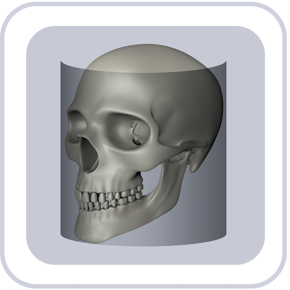
23x16* - SKULL
Ideal for orthodontics, it allow for diagnostics on the whole maxillofacial region.Manuals

PRE-INSTALLATION GUIDE
We have prepared this manual very carefully so that our customers properly prepare the location where the Eagle equipment will be installed. Additional questions can be clarified with our technical team.